


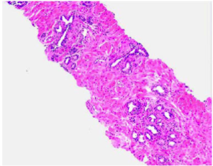
Liver ROIs
Tre projekt beviljade i AIDAs andra ansökningsomgång
I andra ansökningsomgången beviljade AIDAs styrgrupp medel till tre nya projekt. Två av projekten är inom patologi och ett inom radiologi. Den tredje och sista omgången har deadline 22 december 2017.
Decision Support for Classification of Microscopy Images in Digital Pathology Using Deep Learning Applied to Gleason Grading
Anders Heyden, PhD
Lund University, Lund
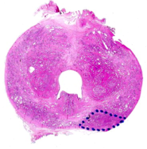
Prostate cancer (PCa) is the second most common malignancy in men worldwide. In Sweden, the incidence is now close to 10 000 new cases per year. Correct identification of the stage and severity of the PCa on histological preparations can help the healthcare specialists predict the outcome of the patient and chose the best treatment options.
The best method of evaluating the severity of prostate cancer in tissue samples is based on the architectural assessment of a histologically prepared tissue specimen (from biopsy or surgery) by an experienced pathologist. The pathologist assigns a “Gleason grade” ranging from 1 (benign) to 5 (severe cancer). This type of cancer severity grading is highly correlated with prognostic outcomes and is the best biomarker in PCa to predict outcome. However, intra-observer differences between pathologists are a major problem. This results in costly repercussions; under-diagnosed patients may become more ill while over-diagnosed patients receive unnecessary treatment, in all decreasing the quality of life and increasing the costs for the healthcare system.
The project aims at developing decision support systems for Gleason scoring of microscopic images of prostate cancer based on deep convolutional neural networks (CNN). We will evaluate the results and obtain feedback on difficult cases from pathologists. The purpose of such a system is to obtain a more reliable estimate of the Gleason score and thus a correct treatment for the patient. The project is based at the Centre for Mathematical Society, Lund University, with close cooperation with the Department of Urology at Lund University Hospital and SECTRA in Linköping.
Interactive Visualization Tools for Verification and Improvement of Deep Neural Network Predictions
Ida-Maria Sintorn, PhD
Vironova AB, Stockholm
Understanding and being able to show what leads to a decision using AI and machine learning methods is important to gain acceptance for their use in clinical diagnostics. The purpose of this project is to adapt and develop interactive visualization tools to explain and test what regions and details in an image are important for the decision of the artificial neural network. Interacting with the visualizations and providing corrections and feedback will further train the neural network for improved performance. The interactive tools will, in addition, allow for convenient hypothesis testing regarding importance of image features by e.g. blocking regions or scales of images or part of a network and seeing the effect on the results.
Verifying what information decision support systems based their decision on is the key for rapid conversion of research results to clinical use. The tools to be developed in this project will hence, not be directly used clinically but rather support and serve as a catalyst for deploying other decision support systems in the clinics. The tools will be evaluated on clinical applications using electron microscopy for kidney diagnoses and light microscopy for cervical cancer screening.
Machine Learning for Automated Measurements of Liver Fat
Magnus Borga, PhD
AMRA AB, Linköping
Non-alcoholic fatty liver disease (NAFLD), a range of diseases characterized by steatosis, is associated with the metabolic syndrome and can lead to advanced fibrosis, cirrhosis, and hepatocellular carcinoma. Non-alcoholic steatohepatitis, a more serious form of NAFLD, is now the single most common cause of liver disease in developed countries and is associated with high mortality. Diagnosis and grading of hepatocellular fat in patients with NAFLD usually requires a liver biopsy and histology. However, as liver biopsy is an expensive, invasive, and painful procedure that is sensitive to sampling variability, the use of MRI as a non-invasive biomarker of liver fat has shown tremendous progress in recent years. Automation of this technology would further reduce costs for clinical use. Measuring fat in the liver is, however, not trivial since larger blood vessels and bile ducts need to be avoided in order to get accurate estimates of the liver fat fraction. Therefore, the aim of this project is to develop an automated method, based on machine learning, for placement of regions of interest (ROI) in which the liver fat can be quantified.
AKTUELLT
Glad sommar önskar vi från Medtech4Health
Efter en intensiv vår är det snart dags för en välbehövlig sommarpaus. Men först ser vi fram emot att ses i Almedalen där vi är en del av mötesplatsen Tillsammans.
SO Västra stärker innovationskraften genom samverkan och erfarenhetsutbyte
Medtech4Healths nod SO Västra har gjort programmet till en naturlig del av innovationsarbetet i Västra Götaland. Här finns en stark samverkan där metoder och erfarenheter delas framgångsrikt.
Nytt verktyg ska bana vägen för smidigare breddinförande av medicinteknik
Först ut från Medtech4Healths strategiska projekt MedImpact är ett instrument för enklare breddinförande av teknik för egenmonitorering. Med instrumentets frågebatteri blir det möjligt att undvika fallgroparna vid ett breddinförande. Instrumentet finns på Medtech Arena.
Nya satsningar ska göra det lättare för små innovativa medicintekniska företag
Regeringen har gett flera nya uppdrag till myndigheter som arbetar med medicinteknik. Målet är att stärka sjukvården och förbättra Sveriges beredskap vid kriser.
NYHETSBREV
Följ nyheter och utlysningar från Medtech4Health - prenumera på vårt nyhetsbrev.