HE_IH_aida
AIDA beviljar medel till Clinical fellowships
I första ansökningsomgången har AIDAs styrgrupp beviljat medel till två clinical felloships. Under projekttiden kommer forskarna att regelbundet vistas i Linköping för att ta del av AIDAs infrastruktur.
Image- and AI-based cytological cancer screening
Eva Ramqvist, MD, PhD
Department of Clinical Pathology/Cytology, Karolinska Univ. Hospital, Solna
This clinical fellowship is aimed at cytological cancer screening, in collaboration with the AIDA decision support project ”Image- and AI-based cytological cancer screening”. Activities include:
- To support the development by detailed manual per-cell annotations and to identify areas with malignancy associated changes, but also to indicate and describe various artefacts and imperfections in the image material. This will significantly increase the ability to develop a robust and well functioning system.
- To introduce cytology as a non-invasive alternative in dentistry, to comply with the World Health Assembly (WHA) goal to increase efforts to reduce oral cancer.
- To organize field studies to evaluate the accuracy and precision of the system, its clinical usefulness and applicability in clinical routine and to introduce the system to the pathology/cytology community.
AI-driven Image Analysis in Digital Pathology
Gordan Maras, ST-läkare
Klinisk patologi och cytologi, Region Gävleborg
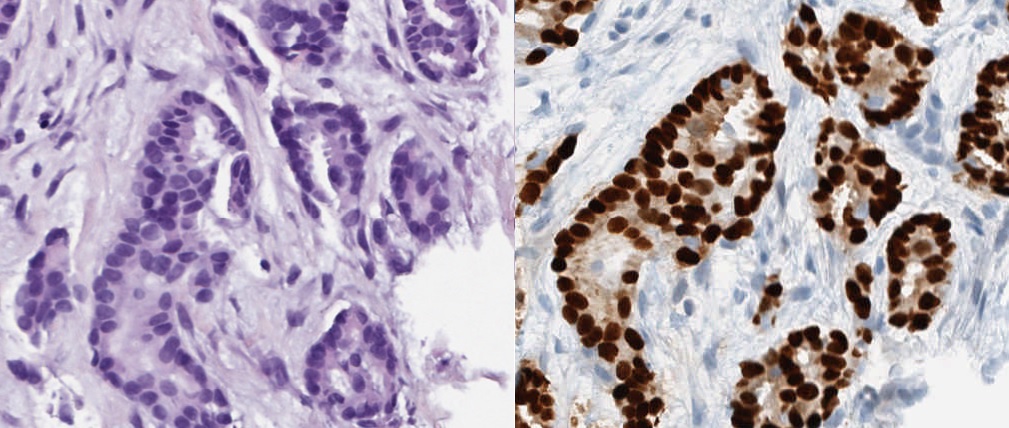
Digital pathology has been gaining attention from both clinical pathologists and technical developers over the last years. One of the interesting possibilities with the digitization of pathology is the use of image analysis. Parallel to the development of digital pathology there have also been an expansion of more advanced technology. An area that has been especially eye-catching is artificial intelligence (AI). The application of AI in digital pathology could potentially create powerful and versatile tools for use in the clinical setting. These could for instance be used for automatic calculation of specific cells or recognition of tumor cells within a lymph node.
In the upcoming project, it would be interesting to investigate the efficiency and the accuracy of AI-driven image analysis when it comes to counting specially stained cells (i.e. hormone positive cells). In addition, it would also be intriguing to look at the speed of the calculations and how these factors may be improved.
These types of tools for the digital pathology would allow for more distinct reporting to various physicians for a better decision making basis which in turn should provide a better and more effective patient care.
AKTUELLT
Ny projektansökningsomgång i AIDA
AIDA är en nationell arena för forskning och innovation inom AI för medicinsk bildanalys. Inom AIDA finns det flera möjligheter till projektstöd.
Vilken roll ges medicintekniken i den nya Life science-strategin?
Under Medicinteknikdagarna sätter Medtech4Health fokus på den nationella Life science-strategin och forsknings- och innovationspropositionen. Vilken plats får medicintekniken, och hur går vi från strategi till verkstad?
Missa inte rabatten till Framtidens hälsa och sjukvård
Medtech4Health är samarbetspartner till evenemanget vilket ger dig 50 procents rabatt på både tvådagars- och endagsbiljetter.
Ny studie kartlägger behovet av samordnad patientsamverkan
Nu finns en ny studie om en nationell samordning och matchning för patientinvolvering i utvecklingen av medicinteknik. Studien har gjorts av Eupati Sverige med finansiering av bland andra Medtech4Health.
NYHETSBREV
Följ nyheter och utlysningar från Medtech4Health - prenumera på vårt nyhetsbrev.


